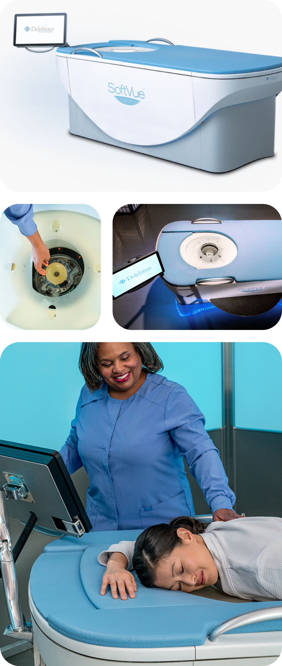
TriAD™ Triple Acoustic Detection Technology
The TriAD™ of Sound
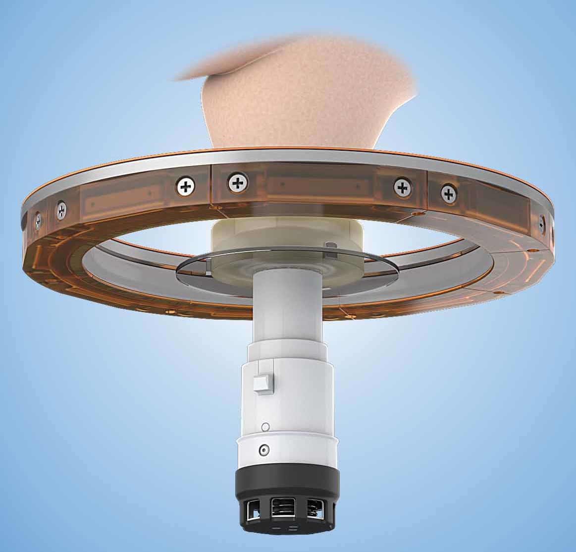
Introducing a singularly unique approach in transducer design, SoftVue incorporates over 2000 transducer elements within a uniform ring configuration featuring TriAD triple acoustic detection. This remarkable imaging capability gathers not only reflected echoes but quantifies the sound speed and attenuation signals transmitted through the breast. The tomographic coronal image presentation delivers an unrivaled view of the structure of breast tissue and depicts tissue attributes of sound speed and attenuation that can assist physicians in distinguishing normal tissue from areas of concern.
Our clinical trial and FDA PMA approval have demonstrated that combining SoftVue with standard mammography increases sensitivity by 20% and specificity by 8% for women with dense breasts.
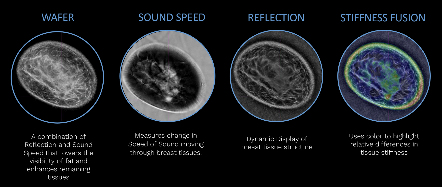
SoftVue™ Offers 4Xs the Imaging Power
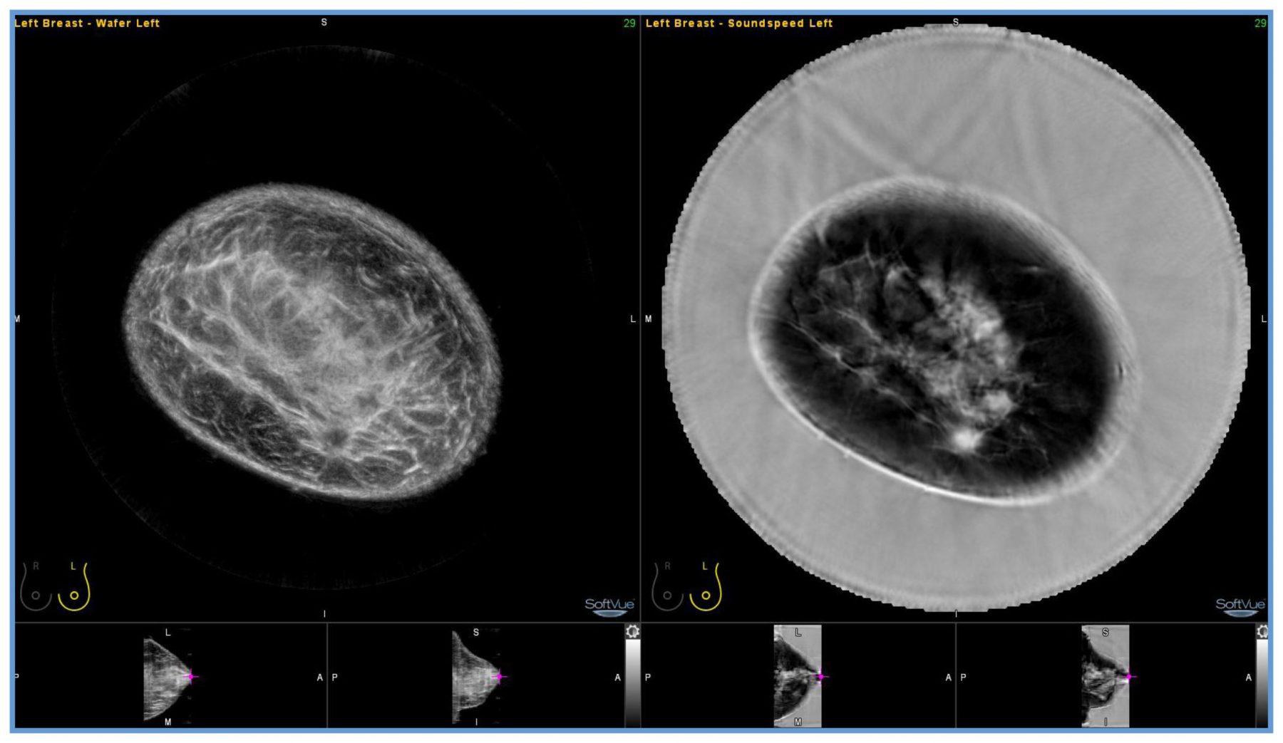
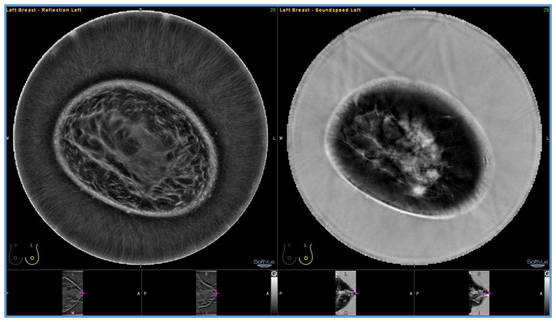
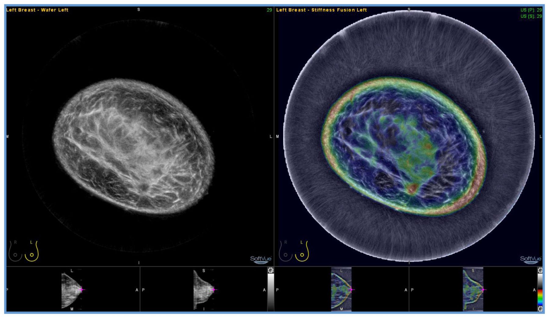
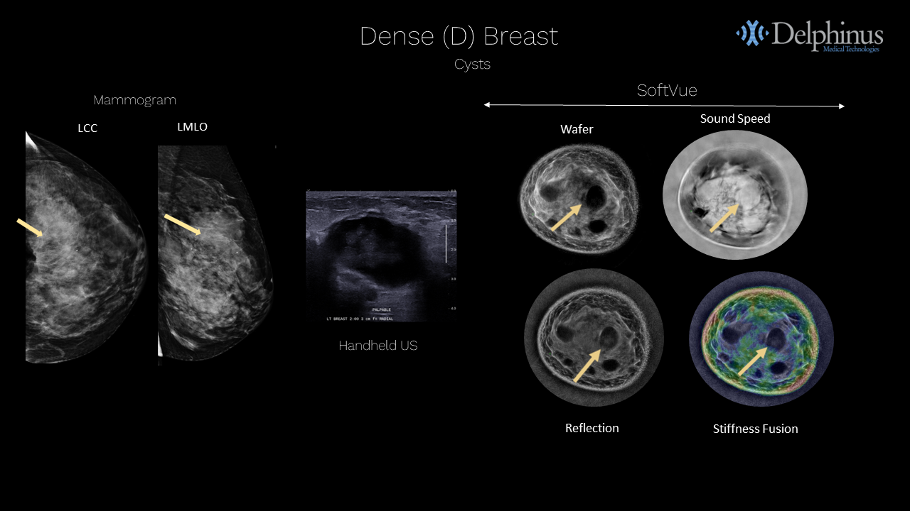
Four volumetric image stacks are provided for interpretation and comparison with other breast imaging studies – Wafer, Sound Speed, Reflection, Stiffness Fusion.
Reflection and sound speed images are included in the final image stack as direct outputs of the image acquisition process.
The backscatter signal of reflection and transmitted signal of sound speed are used to create the Wafer output image that is included in the final image stack.
A Stiffness Fusion image is included in the final image stack and is a combination of the reflection and attenuation signals and replaces the attenuation stack in the final image output. It provide relative differences in tissue stiffness.
These stacks are designed to optimize SoftVue’s tissue specific imaging and provide the inputs for interpretation on the workstation.
SoftVue™ Improves Image Interpretation
SoftVue utilizes traditional ultrasound reflection while improving the clinical information by adding ultrasound transmission to optimize the clinical data.
Ultrasound transmission measures physiologic tissue parameters in sound speed and stiffness, in addition to reflection, a key factor in decreasing the false positive rate.
SoftVue secures anatomical AND physiologic properties of tissue to accurately differentiate cancer from normal tissue or benign disease. These parameters can be used to characterize lesions in a quantitative manner. This quantitative approach is not available in current breast ultrasound systems.
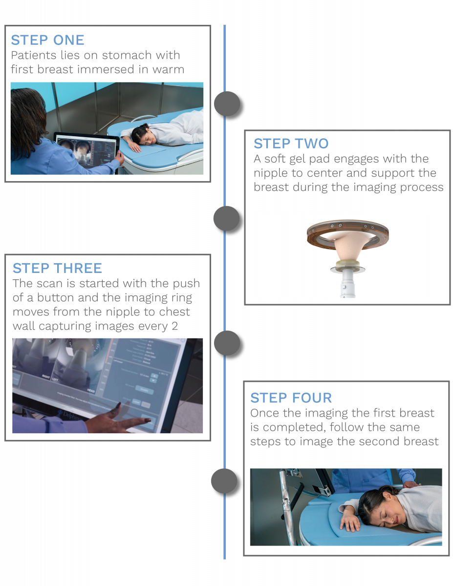
The SoftVue™ Workflow
The SoftVue exam is completed in just a few easy steps with a simplified image review workflow. Following image acquisition, the reflection, sound speed and attenuation sound signals are reconstructed and presented in the coronal plane offering four image sequences for radiologists to use during interpretation in conjunction with a woman’s mammogram.
SoftVue™ Exam Workflow
SoftVue™ Image Sequences
SoftVue™ Image Review Workstation
SoftVue™ Image Review Workflow - Wafer & Sound Speed
SoftVue™ Image Review Workflow - Reflection & Sound Speed
SoftVue™ Image Review Workflow - Wafer & Stiffness Fusion
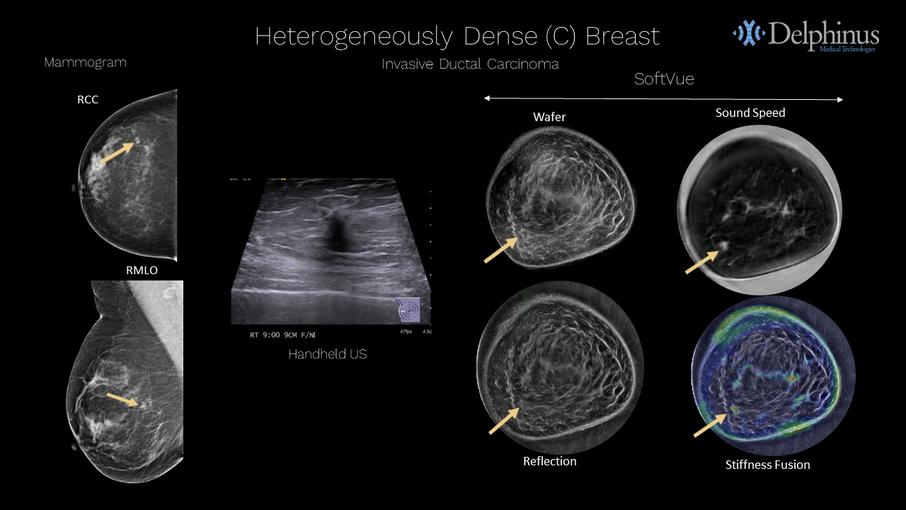
Case Example - Invasive Ductal Carcinoma
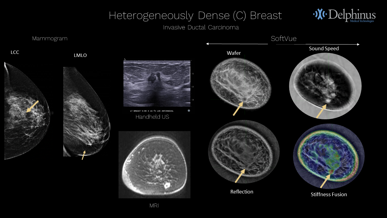
Case Example - Invasive Ductal Carcinoma
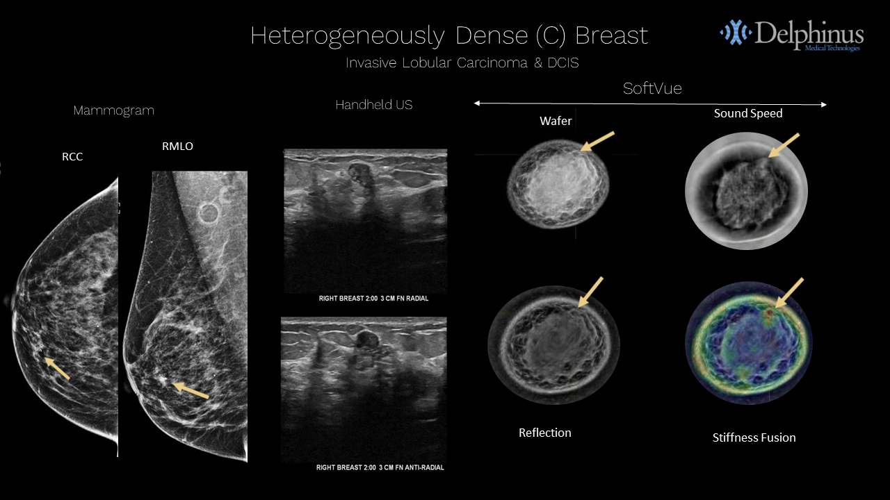
Case Example - Invasive Lobular Carcinoma & DCIS
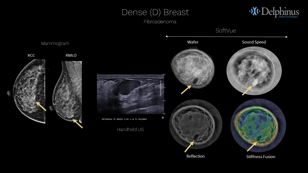
Case Example - Fibroadenoma
Case Example - Cysts

Contact Us for More Information or Demo
Thank you for your interest in a SoftVue™ demo.
Please complete the form below and a Delphinus representative will be in touch.