EXCLUSIVE EVENT FOR HEALTHCARE PROFESSIONALS
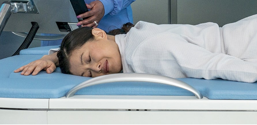
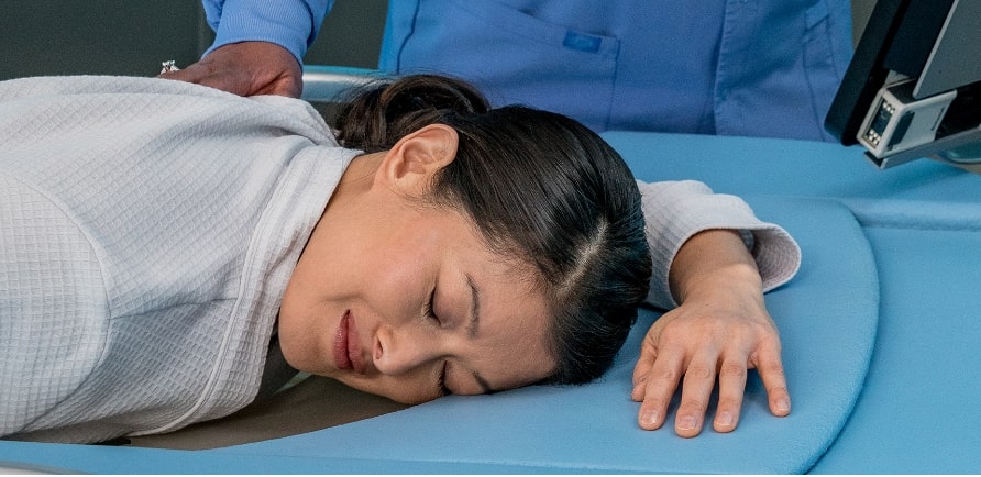
Imagine a dense breast supplemental screening device so COMFORTABLE, EFFICIENT and EFFECTIVE that women recommend it to other women and request it by name.
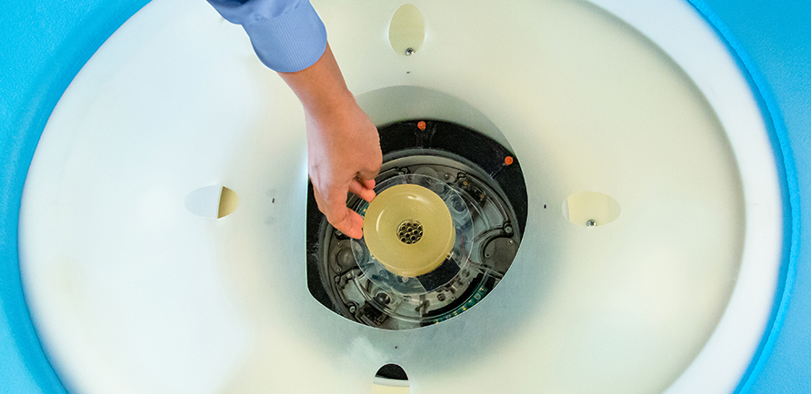
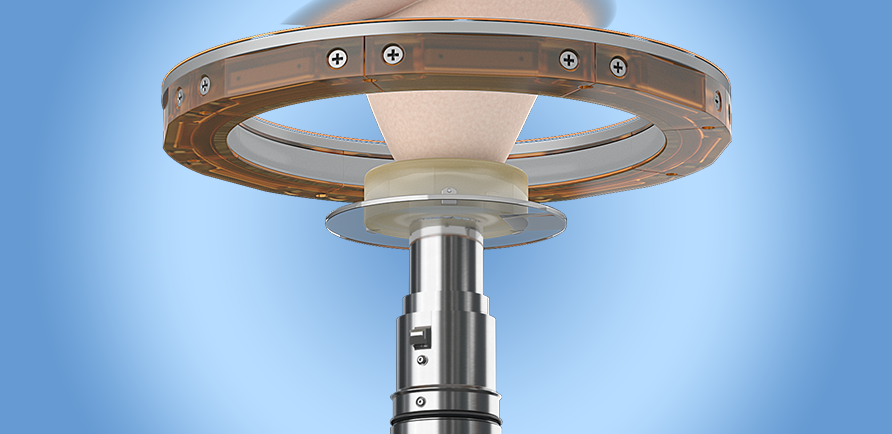
SoftVue™ Breast Tomographic Ultrasound
Exactly what your practice needs to improve cancer detection in your patients with dense breasts and increase patient compliance with dense breast screening guidelines.
SoftVue™ is coming to Austin, TX
95% of women who experienced a SoftVue™ exam
would recommend it to other women
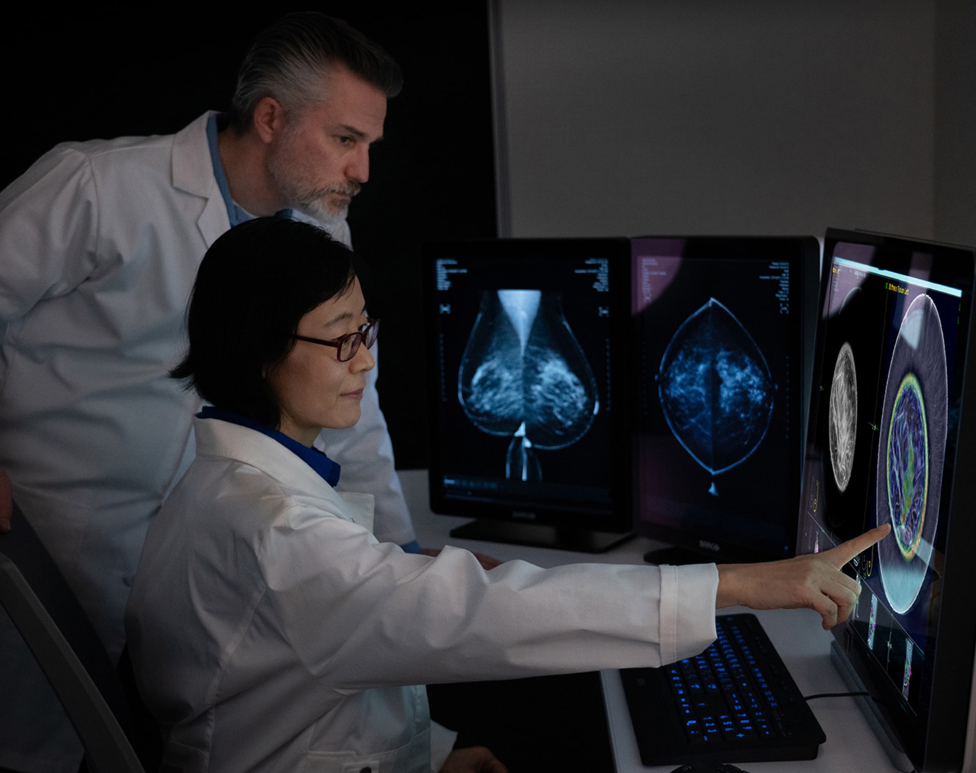
Why SoftVue™ for Dense Breast Screening?
While mammography is highly effective for women with BIRADS a or b breast density, mammography misses cancer in women with BIRADS c or d breast density as both cancer and fibroglandular tissue appear white on mammogram.
SoftVue was developed specifically to address the unmet need for effective dense breast cancer screening.
When combined with mammography:
20%
Identifies up to 20% more cancers with greater accuracy
8%
Increases specificity by 8% at the BIRADS 3 threshold
Register to See SoftVue™
Stop by SoftVue on the Go to see this new technology in action, engage in an image review session and learn more about how SoftVue can set your practice apart from the competition.
Upcoming Locations
Round Rock, Tx
Hosted in partnership with Austin Radiological Association (ARA), SoftVue™ on the Go is coming to Austin! Featuring a live SoftVue scan demo and SoftVue image review session, this is a one-time-only opportunity to learn about one of the industry's most innovative dense breast screening technologies FDA-approved in the past decade. When you couple increased cancer detection with an improved patient experience, you get a technology that will transform your practice. Join us to learn more about the breast ultrasound tomography screening system women will ask for by name, SoftVue.
When:
October 29 & 30 - 60-Minute Educational Sessions including a live SoftVue demo scan and SoftVue image review are offered:
- • 9:00 am – 12 pm
- • 12:00 pm – 1:30 pm
- • 5:00 pm – 6:00 pm
October 31 - 60-Minute Educational Sessions including a live SoftVue demo scan and SoftVue image review offered:
- • 9:00 am – 12 pm
- • 12:00 pm – 1:30 pm
*Registration is required as space is limited.
If you are unable to attend the educational sessions you may come in at your convenience between 9 -11:30 am and 1:30 - 4 pm daily to learn about SoftVue and view a SoftVue image review. However, a live demo may not be available during these hours.
Where:
Embassy Suites270 Bass Pro Dr
Round Rock, TX 78665
Register
Using the calendar, select the date and session you wish to attend SoftVue on the Go and complete the registration process.We look forward to seeing you in Austin!
Want SoftVue available in your area?
Click here and fill out the form and a representative from Delphinus will email you when SoftVue is available in your city.
SoftVue™ Scan Sequence
SoftVue™ Image Sequences
Four volumetric image stacks are provided for interpretation and comparison with other breast imaging studies
Wafer
Wafer
Sound Speed in combination with reflection lowers the visibility of fat and to enhance the remaining tissues.
Sound Speed
Sound Speed
Measurable change in Speed of Sound moving through breast tissue.
Reflection
Reflection
Dynamic display of breast tissue structure.
Stiffness Fusion
Stiffness Fusion
Attenuation in combination with sound speed provides relative differences in tissue stiffness.

Reflection and Sound Speed images are included in the final image stack as direct outputs of the image acquisition process.

The backscatter signal of reflection and transmitted signal of sound speed are used to create the Wafer output image that is included in the final image stack. This sequence suppresses the appearance of fat to accentuate the appearance of tissues and masses.

A Stiffness Fusion image is included in the final image stack and is a combination of the sound speed and attenuation signals and replaces the attenuation stack in the final image output. It provides relative differences in tissue stiffness.

These stacks are designed to optimize SoftVue’s tissue-specific imaging and provide the inputs for interpretation on the workstation.
